Lesson 1 Answers
- (Lesson 1) An ultrasound image is acquired as a series of lines, not all simultaneously.
- (Lesson 1) The echo from the 2 cm deep structure will take 0.02 ms = 20 µs to arrive using the equation in Figure 1, because the wave pulse has to travel 4 cm to and from the structure. The echo from the 4 cm deep structure will take twice as long, 0.04 ms = 40 µs. (1 µs = 1 microsecond = 0.001 ms.)
Lesson 2 Answers
- The figure shows 4 cycles taking place during 0.5 µs = 0.5 x 10⁻⁶ s, so the frequency is 4 cycles/(0.5 x 10-6 s) = 8×10⁶ Hz = 8 MHz.
- In tissue, the wavelength increases compared to the wavelength in air, because the speed of sound in tissue is greater than in air, and the frequency remains the same as the wave travels into the patient. Because wavelength = speed/frequency, increasing speed with constant frequency causes the wavelength to increase. Expressed with equations,
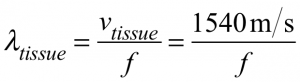 and
and 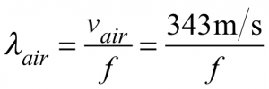 (Lesson 2)
 - Because wavelength = speed/frequency, and speed just depends on the type of tissue, doubling frequency changes the wavelength to half what it was in that same tissue. (Lesson 2)
- To get better resolution (smaller details visible) requires reducing the wavelength; this is achieved by increasing the ultrasound frequency used to acquire the image (see Review Question 2). (Lesson 2)
- Intensity is power per area, so it is reduced due to the loss of energy by absorption in the tissue and due to the spreading out of the beam over a larger area. Because power is just the rate of delivery of energy, only energy loss reduces the power. (Lesson 2)
Lesson 3 Answers
- You should decrease the frequency, as lower frequencies penetrate more deeply. As lower frequencies have longer wavelengths, increasing the size of the smallest structures resolvable, this will potentially degrade the resolution of your image. (Lesson 3)
- A double image could result from refraction of the ultrasound beam as it passes through another structure on its way to the structure of interest. Changing the transducer location and angle might eliminate this artifact. (Lesson 3)
- In the subcostal view (left panel), while not completely perpendicular to the ultrasound beam, the septum is mostly only about 30 degrees off perpendicular and readily generates echos which return to the transducer and can be detected. In contrast, in the apical 4 chamber view (right panel), the very thin atrial septum is parallel to the ultrasound beam and hardly reflects any echos to the transducer. Thus, this structure cannot be well seen. As a result, evaluation of the atrial septum is not recommended from the apical 4 chamber view. (Lesson 3)
Lesson 4 Answers
- (Lesson 4) C is true. Increasing the depth from which echoes can be collected requires increasing the length of time allowed for echoes to return, which is the cycle time. Increasing the cycle time also increases the length of time required to collect a frame (unless the number of lines is reduced, which is not normally done), and therefore decreases the frame rate.
- (Lesson 4) A heart beating 150 times per minute will beat 2.5 times per second, so 5 frames per second corresponds to 2 frames per heartbeat. This is not enough faster than the heartbeat itself to get a good image. Increasing the number of frames per second will improve the image quality by reducing the jerkiness and discontinuity between frames. As a rule of thumb, 20 frames per second gives good quality video, which in this case is 8 frames per heartbeat.
- (Lesson 4) M-mode is ideal for monitoring fast-moving structures such as valves given the high frame rate achievable. In addition, the M-mode display allows tracking a particular structure over time and this can be useful to help identify the structure of interest, such as the endocardial border of the left ventricular posterior wall or the tricuspid annulus.