Estimated time: 15 minutes
Take-home points:
-
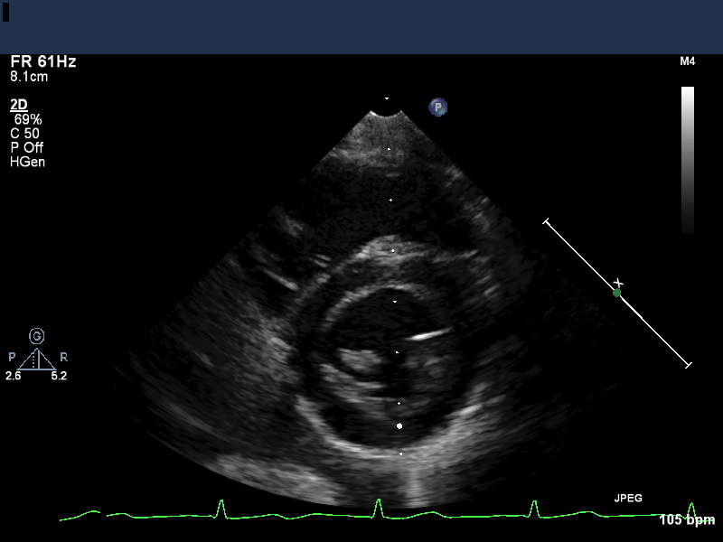
- Echo images can be acquired as single 2D images (still frames, Figure 1), video (a series of frames acquired in rapid succession), or “M-mode” (the signal from a single line repeated very rapidly in order to monitor motion of particular structures).
- Frame rate is an important aspect of video quality, particularly for pediatric patients with higher heart rates
Ultrasound pulse timing and duration
-
- The ultrasound transducer produces the ultrasound pulses and detects reflected ultrasound (echoes), but can’t do both simultaneously. Consequently, the pulses have to be brief enough and spaced enough in time to receive the echoes between pulses.
- Most of the time, the transducer is receiving (“listening”) rather than transmitting ultrasound. This is expressed as a parameter known as the “duty factor,” which is the percentage of the time that the transducer is transmitting. Duty factor is typically on the order of 0.1%.
- The time from the start of one pulse to the start of the next is called the cycle length, Tcycle. This is also the time required to obtain one line of an ultrasound image. The inverse of cycle length is the pulse repetition frequency (PRF). This is different from the fundamental frequency of the wave f, but has the same units.
- The ultrasound pulse encounters several reflecting structures as it travels into the patient. Each structure reflects part of the pulse and transmits part, so multiple echoes are received. The lower intensity reaching deeper structures means those echoes may be weaker. The weaker signals from deeper structures are compensated for by the Time Gain Controls (TGCs), where in the machine augments (increases gain) of the signals from later (and thus deeper) echos.
M-mode imaging
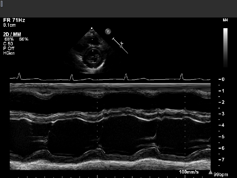
M-mode (or “motion-mode”) imaging is an implementation of cardiac ultrasound in which the data from a single scan line is plotted versus time, as shown in Figure 2. Currently, the 2D image shown at the top of the figure is used as a guide to select the single scan line of interest, but historically, M-mode imaging was developed before 2D imaging and was adjusted by the operator based on the visualized M-mode tracing. The acquisition of 2D images is discussed below.
-
- Because only one line is being acquired, that line can be sampled very frequently, limited only by the time needed for the ultrasound to travel to the required depth and back (the cycle length Tcycle).
- In practice, M-mode sampling rates are typically about 1800 times/second.
- This high sampling rate facilitates accurate identification of continuously moving structures such as the ventricular endocardium or the tricuspid annular plane.
- We typically use M-mode for accurate measurement of left ventricular dimensions or tricuspid annular plane systolic excursion (TAPSE). Other applications include assessment of rapid intracardiac motion of valve leaflets or vegetations.

2D echocardiography
A single still image (“frame”) of an echocardiographic study is assembled from many individual lines that radiate out from the transducer (Fig. 3).
-
- After a set of echoes is obtained from a given line, the next pulse is sent in a slightly different direction to obtain the next line.
- The resulting image is wedge-shaped, with the point of the wedge at the location nearest the ultrasound transducer (see Figure 1).
- Typically 128 lines are collected for each image.
Video imaging considerations
The frame rate is the number of individual still images (“frames”) per second that make up the video; higher frame rates give better quality video, with more continuity from image to image. Acquisition parameters need to be chosen so that the frame rate is adequate to capture the information desired. These acquisition parameters will be discussed further in Module 2.
-
- A frame rate of 20 frames/s or more is normally adequate for the video to appear essentially continuous and reveal all necessary dynamics. This requires each frame to be collected in 1/20 of a second = 50 ms.
- The heart rate of a newborn is typically 150 beats/minute = 2.5 beats/second, so a frame rate of 3 frames per second would provide roughly one frame per heartbeat. The image of the heart would be significantly distorted because different lines would be collected at different times in the contractile cycle.
To capture an entire frame consisting of 128 lines in 50 ms, each line must be acquired in 0.39 ms. Each line corresponds to the transducer sending a single pulse and then receiving its echoes before sending the next pulse.
-
- For a cycle length of 0.39 ms, the maximum depth d of structures from which echoes can be collected is found using the wave speed v and cycle length T. The pulse must travel a distance 2d for the echo to be collected, so the maximum depth satisfies 2d = vT and thus d =vT/2.
- Substituting 1540 m/s as the speed of sound in tissue, and 0.39 ms as the cycle time, echoes could be collected from depths as great as 0.30 m or 30 cm — which approximately equals the maximum ultrasound penetration depth. So with these parameters, there is plenty of time to collect echoes from physiologically relevant depths.
- To increase the frame rate, the image depth can be decreased, which reduces the cycle time. This and other adjustments to improve frame rate are further discussed in Module 2.
Review questions
- Suppose you increase the depth setting in the course of an echo study. Which of the following statements is true?
A. The echo machine will increase the cycle time and you will also see an increase in your frame rate.
B. The echo machine will decrease the cycle time and you will also see an increase in your frame rate.
C. The echo machine will increase the cycle time and you will also see a decrease in your frame rate.
D. The echo machine will decrease the cycle time and you will also see a decrease in your frame rate
(Answer) - Could you obtain clear video of a pediatric heart beating at 150 beats per minute with a video frame rate of 5 frames per second? If you wanted to improve the quality of the video, would you try to achieve a larger or smaller number of frames per second ? (Answer)
- Under what circumstances would you use M-mode imaging? (Answer)